Experienced Brain Injury Attorneys Who Understand Your Loss
Brain injury deserves specific mention because it represents damage to our very being. Whether mild or severe, brain injury is always serious because it has markedly altered an individual’s ability to contribute to life. This is why we focus so much on this sensitive area of personal injury law.
As an experienced brain injury attorney based in Jackson, Wyoming, Bob Schuster has represented victims of traumatic brain injury his entire career. These cases often result from physicians paying too little attention to fetal heart rate in the hours before a baby’s birth, a child breathing carbon monoxide (a colorless, odorless gas) in her bed, or a traumatic blow to a person’s skull when struck in an unfortunate collision. They are always tragic and always heartbreaking.
Whether you are seeking a birth injury attorney, a carbon monoxide lawsuit or representation following a car accident, we will ensure that your case is backed by an expert resource team and that you receive the personal attention you deserve during this difficult period. As a member of the American Association for Justice‘s Traumatic Brain Injury trial lawyers group, Bob Schuster is a trusted resource on the latest legal strategies and tactics in this practice area. Contact him today for a consultation on your case.
Brain Injury Facts
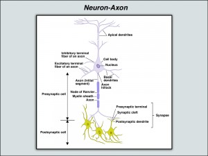
Carbon monoxide poisoning is just one example of the cause of brain injuries. It can result in severe injury to multiple areas or structures of the brain, including the white matter and deep gray matter structures such as the basal ganglia (particularly the globus pallidus and the putamen — both of which are located within the basal ganglia) and the hippocampus. The white matter is composed primarily of filament-like fibers called “axons” that transmit nerve messages from cell to cell and, accordingly, damage to the white matter can cause profound and generalized impairment. The brain and its structures are extremely susceptible to damage from carbon monoxide. Because it is an organ essential to life, the brain is given an oxygen priority by our bodies. If brain cells do not receive oxygen, they will die and, in contrast to other cells, will not be replaced. The potential for damage is profound because the smallest cell within the brain requires oxygen — which means that the arterial system becomes microscopic in its elaboration to reach each of the cells in the brain.
If that arterial system is unable to transport oxygen to the cells because the hemoglobin has been covered by carbon monoxide molecules rather than life-giving oxygen molecules, brain cells will die — and will die in all areas of the brain, including its farthest and deepest reaches. Carbon monoxide is particularly lethal to brain tissue; however, not only because it starves the cells of oxygen but it also destroys brain structures because it is a neurotoxin.
Some of the areas of the brain impacted by carbon monoxide poisoning are described below:
Cerebrum
The cerebrum is the term used to describe the major structures of the brain, excluding the lower structures that connect to the spinal cord: the cerebellum, the mid-brain, the pons, and the medula oblongata.
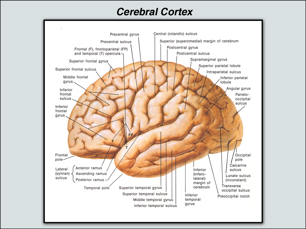
Cerebral Cortex
The cerebral cortex is the thin (about 3 mm in depth) layer or mantle of gray matter covering the surface of each cerebral hemisphere and it is folded into gyri (rounded, protruding undulations) separated by sulci (fissures or valleys). Being gray matter, it is composed of neuronal structures and is an area that is significant for higher mental functions, general movement, perception, and behavioral reactions — and the integration and association of these functions. And, as gray matter, it is dependent on the integrity of the axonal structures comprising the white matter, because it is the axons over which the messages and nerve transmissions travel. The term “cortical” refers to the cortex; whereas, “subcortical” describes those structures that are beneath the cortex and can include both gray matter structures as well as white matter fiber tracts.
Frontal Lobes
The frontal lobes are the front portion of the two hemispheres of the brain.
Temporal Lobes
The right and left temporal lobes of the cerebrum are located on the side and at the base of the brain; in front of the occipital lobes and below the parietal and frontal lobes.
White Matter
The white matter of the brain is composed mainly of axons — the filament-like myelinated fibers that carry nerve impulses between neurons. Neurons are the nerve cells involved with neural processing and which are located in the gray matter; their function is to analyze and transmit information. The axons that comprise the white matter are responsible for the actual transmission of information while the neuron bodies in the gray matter are responsible for information processing. The white matter forms the bulk of the deep parts of the brain. Because the axons transmit nerve messages from cell to cell, damage to the white matter can cause profound and generalized impairment.
Ventricles
The ventricles are four connected cavities within the brain that are filled with cerebrospinal fluid (CSF). The two larger structures — the right and the left ventricles — are located in the respective hemispheres of the brain within the frontal, occipital, and temporal lobes. The third ventricle is smaller and is positioned between the two lateral ventricles — with the cerebrospinal fluid flowing from each through a channel called the interventricular foramen. The fourth ventricle is positioned even lower — and the CSF moves from the third ventricle through the narrow cerebral aqueduct of Sylvius. The cerebrospinal fluid — in the ventricles as well as in the subarchnoid spaces and the spinal canal — acts as a liquid buffer or cushion, absorbing and distributing forces and providing the brain with a measure of resilience. The volume of CSF can vary, thereby permitting the regulation of the total capacity of the cranium. This feature of ventricular activity has particular relevance to brain injury that causes atrophy. The ventricles can increase in size, filling the space that used to be occupied by the healthy brain tissue before its damage, death, and atrophy. Accordingly, enlargement of the ventricular spaces (dilation) can be clinically important in assessing the extent of atrophy and brain damage. The dilation can also be noted with other spaces, in addition to the ventricles. For instance, the perivascular space — the area surrounding blood vessels in the brain (also known as Virchow-Robin spaces) — can be dilated, indicating atrophy in the adjacent tissue structures.
Basal Ganglia
The basal ganglia is a collection of nuclei or structures embedded in the lower parts of each cerebral hemisphere. It includes the lenticular nucleus (which is comprised of the putamen and globus pallidus), the caudate nucleus, the subthalamic nucleus, and the substantia nigra. The basal ganglia is extensively linked to the cerebral cortex and is associated with complex cognitive behaviors (including learning, integrative cognitive functions, memory, emotional functions, and control or regulation of behavior) as well as regulation of motor functions, including limb and eye movements. The basal ganglia are responsible for retrieval processes — the ability to retrieve information stored in the brain — as well as procedural memory — the memory associated with the knowledge to do a task. (Bigler and Clement, Diagnostic Clinical Neuropsychology at 44. 3d Ed.) These structures are vulnerable to damage from carbon monoxide poisoning.
Globus Pallidus
The globus pallidus is part of the basal ganglia, comprising, with the putamen, the structure known as the lenticular nucleus. It is considered to be an “output nucleus” of the basal ganglia, linking with the caudate nucleus to form the corpus striatum and constituting the functional part of the basal ganglia. The globus pallidus projects its “outputs” to the other parts of the brain, including the anterior and lateral nuclei of the thalamus, the subthalamic nucleus, and the midbrain tegmental areas. Accordingly, damage to the globus pallidus can severely impact the functioning of the basal ganglia, and thereby can impact cognitive as well as motor abilities.
Putamen
The putamen is also part of the basal ganglia, being one of the components (with the globus pallidus) of the lenticular nucleus. It receives inputs from the motor and sensory areas of the brain’s cortex and then transmits them to the globus pallidus, the “output nucleus” of the basal ganglia. Damage to the putamen, therefore, can result in serious motor and sensory limitations.
Hippocampus
The hippocampus — from the Greek word for “sea horse” — is a curved structure deep in the cerebral cortex and extending the length of the floor of the lateral ventricle’s temporal horn. It has substantial connections with the hypothalamus — which is involved with the regulation of emotional behavior — and with the temporal lobes — both of which are involved with learning and memory. The hippocampus, for instance, has been shown to have a significant role in the ability to learn new information and to retain it in long-term memory.
Additionally, the hippocampus is associated with regulation of the autonomic nervous system — that functioning that occurs involuntarily and without our conscious direction. As one of the major structures of the limbic system of the brain, the hippocampus is associated with functions such as reasoning, problem-solving, and the initiation and inhibition of behaviors. It has been noted that “the components and connections of the limbic system contain most of the elements that define individual personality, cognitive style, and patterns of behavior.” Bigler and Clement, Diagnostic Clinical Neuropsychology at 14 (3d Ed.)
It is worthwhile — in assessing the impact of brain injury to specific parts of the brain — to understand that there are different types of memory, including declarative (memory of facts and events) and procedural (memory of how to do something). The hippocampus, for instance, is crucial to the consolidation and functioning of declarative memory, whereas it appears to be less involved with procedural memory. (Bigler and Clement at 42-3).
It also is important to short-term memory processing (Bigler and Clement at 44) and emotional processing (Gazzaniga et al, Cognitive Neuroscience: The Biology of the Mind at 85. 2nd Ed.) Following carbon monoxide poisoning, the victim can suffer bilateral hippocampal atrophy that may not develop for weeks or longer after the exposure.
Corpus Callosum
The corpus callosum is the brain’s largest fiber track pathway. It interconnects the two cerebral hemispheres and is divided, front to back, into four parts: the rostrum, the genu, the body, and the splenium. It is a fundamental structure, and its fiber tracts are essential to the interhemispheric exchange of brain signals, including the support of memory, learning, and sensory functions of the brain.
Related: Brain Injury Legal News


